Ear Pathologies and Audiograms Part 4: Meniere’s Disease

For this blog we will be looking at Meniere’s disease. Remember audiograms will differ depending where they are along the pathology pathway. Meniere’s causes a progressive hearing loss in sufferers. I will try and give examples of audiograms early on the disease pathway and later on when the hearing loss has progressed. Once again these are examples, of audiograms taken from clients. Meniere’s disease is very individualistic and the how the different symptoms present and how they affect the sufferer will vary from individual to individual. Because of how complicated Meniere’s can be this will be a very long blog!

Meniere’s

According to the American National Institute on Deafness and Other Communication Disorders ‘Meniere’s disease is a disorder of the inner ear that causes severe dizziness (vertigo), ringing in the ears (tinnitus), hearing loss, and a feeling of fullness or congestion in the ear. Meniere’s disease usually affects only one ear, but in 15% to 25% of people with the disorder, both ears may be affected. Attacks of dizziness may come on suddenly or after a short period of tinnitus or muffled hearing. Some people have single attacks of dizziness separated by long periods of time. Others may experience many attacks close together over several days. Some people with Ménière’s disease have vertigo so extreme that they lose their balance and fall’. https://www.nidcd.nih.gov/health/menieres-disease (I have even seen cases where the individual is unable to get out of bed and function normally, the vertigo is so persistent and severe).

We rarely see it in individuals under 18 years of age. It is most likely to occur in adults between the ages of 40-60 years of age. No one is quite sure what causes Meniere’s disease. Where it occurs in more than one family member it is thought genetic variations may play a part in the origin. About 7-10% of those affected have a family history of the condition.  Incidence of Meniere’s is between 1:1000 and 1:2000 of the population. (Meniere’s UK)

What happens in the ear

When you see a picture of the cochlear you will see that it contains a snail like bit called the cochlea and two upwards facing semi-circular canals these are the organs of balance NIDCD explains it as follows ‘(the semi circular canals and otolithic organs) and the cochlea and has two sections: the bony labyrinth and the membranous labyrinth. The membranous labyrinth is filled with a fluid called endolymph that stimulates receptors in the balance organs as the body moves. The receptors then send signals to the brain about the body’s position and movement. In the cochlea, the fluid is compressed in response to sound vibrations. This stimulates sensory cells that send signals to the brain. In people with Meniere’s disease, a build up of endolymph in the labyrinth called endolymphatic hydrops disrupts normal balance and hearing signals between the inner ear and the brain. This disruption is also associated with vertigo and other Meniere’s disease symptoms’ https://www.nidcd.nih.gov/health/menieres-disease.

We think this endolymphatic hydrops causes an increase in the pressure in the endolymphatic space. It is quite likely that this increased pressure is what causes the fullness and reduced hearing.  Meniere’s UK explains it quite nicely ‘The sudden release in pressure accounts for the sudden attacks of vertigo. Repeated episodes of high pressure and sudden releases of that pressure damage the delicate structures of the inner ear and the balance structures of the semi-circular canals. This cumulative damage results in a decline in hearing levels over time.’

Symptoms of Meniere’s

Meniere’s is very individualistic; symptoms vary between people and over time. The main symptom is attacks of vertigo with nausea and vomiting. These attacks can last from a few minutes to 24 hours (or longer in some people).  During the attack people may also suffer from tinnitus, a feeling of fullness in the affected ear and hearing loss. After the attack the hearing may improve, but generally never to where it was previously. The tinnitus and fullness may go.

For some tinnitus is there all the time with Meniere’s but worsens during the attack. As Meniere’s disease effects the low frequencies initially, many sufferers indicate that the tinnitus sound is often more low frequency in pitch like a rumbling.

Periods of remission between attacks can vary from days to months or even years. For some the vertigo attacks can be severely debilitating causing them to be bed bound; as just raising their head will make the dizziness worse and induce nausea and vomiting. Whilst other individuals may be less affected. As it progresses the vertigo may become less severe. In the later stages tinnitus is often more prominent and the fluctuating hearing loss worsens. Some individuals with Meniere’s will not just have a significant hearing loss but also find that what they are hearing, even with hearing aids in, is severally distorted and may also suffer from sound tolerance issues whilst others may not have this issue at all.

Some people use some of these symptoms as forewarning as to when an attack is about to occur. I have had many clients over the years say that a sudden change in the ear fullness or increase in the tinnitus alerts them to the fact that an attack is imminent.

Lastly whilst stress is not a trigger to Meniere’s we know that it can increase the frequency and intensity of attacks.

Diagnosing Meniere’s disease

GPs cannot diagnose Meniere’s disease. Only Ear, Nose and Throat Surgeons/Otolaryngologists are able to diagnose the disorder after evaluating all the information  from testing and medical history. There is no one test that allows for diagnosis. The client’s history, hearing tests, speech understanding tests, blood tests, MRI scans and vestibular testing procedures which check the balance mechanism will be used in order to get a diagnosis. For further information on these visit Visiting ENT, diagnosis and testing | Ménière’s Society

Intervention Options

Hearing Aids

Hearing aids can be helpful for suffers, and many will find it improves their hearing. However, the sufferer needs to have a clear understanding of the limitations also. Speech in noise understanding will still be difficult, and hearing aids will need to be adjusted as the hearing loss changes and in the later stages, discrimination becomes further impeded. You may wish to consider a CROS aid for the affected Meniere’s ear, particularly in the mid to late stages of the disease. For those with hyperacusis issues or severe distortion then hearing aids may be very limited in their benefit or may not work at all for that ear. Hearing aids may also help with the tinnitus. (remember tinnitus therapy is also available on the NHS if this is needed). I would always suggest trying a hearing aid, but both of you must have a clear understanding and discussion about the limitations and benefits and the issues that may occur.

Surgery

Where the balance/vertigo is severely affecting the client’s quality of life is then surgical intervention may be the only option. There are different options here such as intratympanic injections and endolymphatic sac surgery. Surgery | Ménière’s Society

Drugs/Therapy

Medications can be used to help manage vertigo symptoms Medication | Ménière’s Society. Some people also find adjusting their diet and reducing their salt intake helps with symptoms.  Also managing stress is very important in helping control the symptoms. Controlling Your_Symptoms_(Nov_2023).pdf. There are also balance rehabilitation exercises that help you manage your symptoms Balancing_Retraining_(Nov_2023).pdf

Audiograms

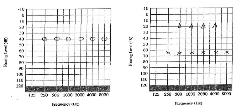
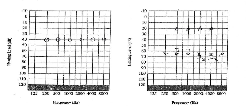
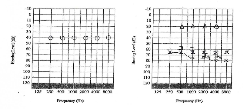
The “classic” picture of Meniere’s hearing loss is that there is a fluctuating, gradually progressive sensorineural hearing loss. It starts with low frequency  hearing loss, and eventually progresses to become “peaked” (both low and high tone), and then flat. Audiograms and description from Hearing loss in Meniere’s disease

Early Meniere’s Disease Left ear : Low Freq are reduced

Mid stage Meniere’s Disease Right Ear: Low Freq worse but flattening out

Late-stage Meniere’s Disease Right ear: Hearing aid likely to be ineffective CROS/BICROS better option

Introducing Louise Hart

Louise Hart joins Tracy James to deliver tinnitus and hyperacusis services to adults and children from October 2020. She will also be providing her expertise in our training courses delivered to provide British Society of Audiology Certificates in impression-taking, hearing surveillance and audiometry and tympanometry.

Louise says ‘ I am excited to be providing more independent work, alongside my NHS work. I really enjoy training and have pride in helping individuals enable their skills to the recognised standard of the British Society of Audiology. We will be training GPs, teachers of the deaf, assistant audiologists and any professional who works in the hearing industry, and I look forward to meeting our new delegates at the next course in Newbury.’

‘With regards to tinnitus, I will be providing the only independent tinnitus and hyperacusis management service in West Berkshire at Tracy James Hearing. We know the earlier we intervene in helping people manage their tinnitus and hyperacusis the more successful the outcome for them’

Despite research on drug or physical interventions on tinnitus, at present none seem to consistently reduce tinnitus well enough; this is why management techniques are used to combat tinnitus.  For over 20 years chronic pain sufferers have successfully been using cognitive behavioural techniques to manage pain, and we now have more studies showing the same success with tinnitus.

Louise will tailor a programme to you to help your tinnitus and/or hyperacusis; these can be provided face to face or through video consultation. For further information on tinnitus and or to book an appointment, go to Tracy’s website tjhearing.co.uk.

For further information on audiology training courses, go to tjaudiology.com


OTIS Simulation software

Audiometry Simulation Software


Otis the virtual patient (from Innoforce.com) can be used to practise basic audiometry and tympanometry and masking. We use it in our classes and its a great way of developing your skills in audiometry without having the pressure of time or the client’s ability to focus, while getting used to the controls and techniques required. If you’re learning how to carry out masking, it is an invaluable learning and evaluation tool and you don’t need to rely on your supervisor to explain results.


Otis is a little bit like a real person, for example, your client can fall asleep if you take too long or shout if you present sounds that are too loud. You can view your otoscopy, history and tymp data as part of the assessment. There is reference data for masking and symbols. You have real-time evaluation and assessment of your procedure as you do the audiometry – it tracks your accuracy, time and errors. You can also progress from easier audiograms, to more difficult cases.


For further information please see our course guide to Audiometry & Tympanometry

Rule 3 explained? Masking Training with Tracy

In this post I would like to share some thoughts about rule 3.

This is the rule that many people scratch their head over, mainly because it can be difficult to see, but also because it doesn’t come up that often (and generally only in more complex cases). Therefore, if you’re used to testing routine cases you may not have to think about rule 3 very much. That’s why it’s good to refresh your knowledge so that you don’t miss it when it does come along. Completing rule 3 means that your hearing test will be more accurate; the end result may affect your diagnosis and also your hearing aid prescription.

How does Rule 1 differ to Rule 3 in masking?

We know that Rule 3 is only needed when Rule 1 has not been carried out and that both involve air conduction masking. But why?

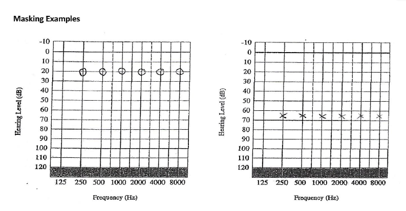
Masking is carried out when there is a difference of 40dB between cochleae when using headphones. When we look at Rule 1 the difference between the two cochleae are obvious:

Masking 1

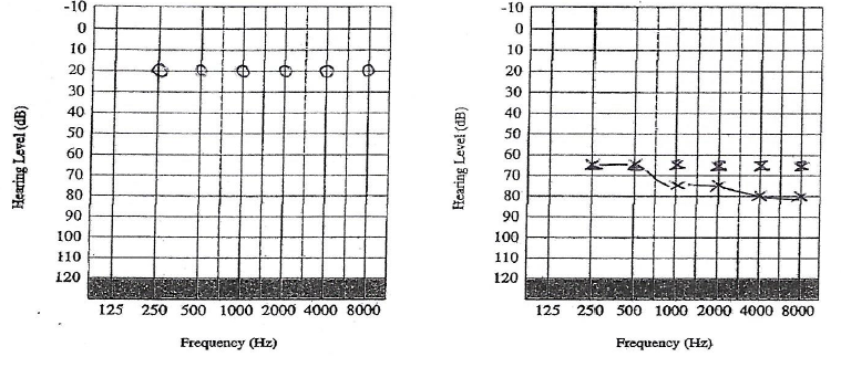
We mask the air conduction, and find the following:

Masking 2

Rule 1 was carried out at all the frequencies as there was a difference of >40 dB  at all the frequencies between the 2 ears. Headphones were used and the left ear was the test ear, and the right ear was the non-test ear, to be masked.

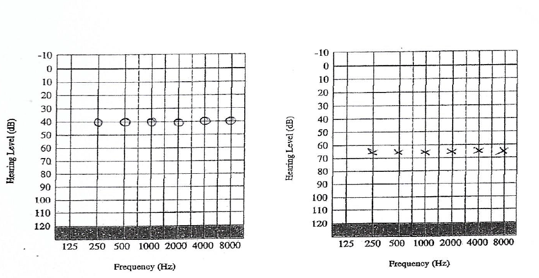
However, the difference between the two cochleae can be hidden when the non-test ear has a conductive component. Say for example the same patient as above comes back a few weeks later with a hideous cold that has affected his right ear.

Masking 3

There was no need to do air conduction masking in this scenario (Rule 1 not required).

Bone conduction was carried out on the worse ear:

Masking 4

As the there was an air bone gap of >10dB between the air and bone conduction thresholds (Rule 2), bone conduction masking needed to be carried out at 0.5, 1, 2 and 4 kHZ. The left ear was the test ear using bone conduction, and the right ear was the non-test ear, to be masked.

Once bone conduction was completed, we could see an asymmetry once again:

Masking 5

In this case we could only see that the right cochlea was >40dB better than the left cochlea because bone conduction had been completed. So we went back to using headphones again and masked the air conduction, with the masking noise in the right ear (non-test ear), and retested the hearing thresholds on the left ear (test ear). This revealed the following:

Masking 6

Moderate to severe sensorineural hearing loss on the left. Mild conductive hearing loss on the right.

Rule 3 explained? (Some tips):

  • Rule 3 is usually required because the non-test ear has a conductive component
  • The asymmetry that is present between the 2 cochleae (as you would visibly see in Rule 1) is hidden until you carry out the bone conduction. That is why you only carry out Rule 3 if Rule 1 has not been carried out.
  • Rule 3 is often missed because the BC that relates to the better ear (non-test ear) is recorded on the side on which the BC is placed (worse ear). Therefore you have to think to which ear the not-masked BC belongs to.
  • Although you check the BC to ascertain an asymmetry, you don’t use the BC in your masking procedure – you are masking the AC thresholds only.
  • At frequencies where no b-c thresholds have been measured – if there is a possibility that a-c threshold at these frequencies (including 250 Hz and 8000 Hz) are not the true thresholds, they should be masked.

If you have any questions please send an email to [email protected] and either myself or Louise will get back to you

Tracy James visits Estonia

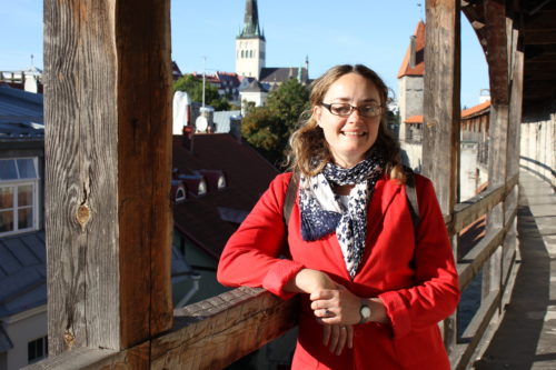
Tracy James of TJ Audiology delivered a 3 day BSA certificate training course in Audiometry and Tympanometry at Medivar in Tallin, Estonia. Tallinn was fantastic, with a beautiful old town; cobbled streets, 14th Century buildings and lots of history. Estonians are also very exotic with only 1 million inhabitants. For those of you that don’t know – Estonians speak Estonian which has a lovely sing song lilt to it.

The training course was delivered to 5 employees in English and I was amazed at how those on the course (and Europeans in general) were able to use English in their day to day lives. Everyone took great assurance in having lots of practice time and as the group was small, there was lots of time to make sure that questions could be answered. Some of what we covered included anatomy and physiology, otoscopy images (possible abnormalities we may come across), contraindications, physics of sound as well as the practical procedure.

Feedback included: “Very thorough and in-depth. Informative, professional and gave consistent support and feedback to the practical assessments as well as to how we were progressing in general. Pace was perfect.”

I was pleased to have visited both Estonia and Medivar and to learn more about Estonian culture as well as Audiology Services. Medivar is a European distributor of health equipment, including Audiology equipment and supplies. I found the people I worked with there very professional and I was very well looked after. Thank you very much!


Tallinn Audiology Training Estonia Audiology Town Walls Estonia Audiology Training


HSE and Hearing Surveillance – Industrial Audiometry UK

The HSE indicates there is variation in practice and standard across practitioners in Hearing Surveillance Programmes. Does your occupational health worker attend a British Society of Audiology accredited course?

Tracy James MSc

The Health and Safety Executive (2013) carried out visits and questionnaires across a range of health surveillance programmes across the UK so that current practices across teams and individuals could be evaluated. Their key findings included that there was variation in practice across practitioners and health surveillance programmes. In particular, they noted that:

 ‘….some practitioners probably did not adopt sufficient measures to exclude the effects of prior exposure to noise and background noise when the test was being conducted’

…and with regards to training competency they noted:

If practitioners had undergone appropriate training there is an assumption that they would be competent to undertake otoscopy, but it was found that otoscopy was not always carried out before testing’

HSE (2013). Current Practice in Health Surveillance for Noise, pp iii

The Health and Safety Executive (HSE) outlines clearly what is required with regards to a health and surveillance programme under the Guidance on Regulations ‘Controlling Noise at Work’ 2005. In Appendix 5 it reports that a training syllabus for industrial audiometricians has been prepared by the British Society of Audiology (BSA) which has accredited a number of courses. The document is available https://www.thebsa.org.uk/wp-content/uploads/2023/10/OD104-65-Surveillance-Audiometry.pdf

Why choose a BSA accredited course? In order to be accredited, the provider has submitted the course content and assessment material to experts in the field to ensure that the training is accurate and according the recommended procedures published by the BSA. It also ensures that the minimum training guidelines are fulfilled and that the trainer has a suitable background knowledge/qualifications for providing the training. A representative of the BSA will also visit the course provider during their training programme.  In order to remain accredited, course providers are required to resubmit their training material every 3 years to ensure the course is up to date.

BSA industrial audiometry courses are designed to enable occupational health workers to:

  • Undertake a brief subject interview and/or administer a questionnaire regarding otological and noise history
  • Perform otoscopy and pure-tone a-c threshold audiometry without masking, both in accordance with BSA recommended procedures. Audiometry may be manual or automated
  • Interpret results and classify them in accordance with HSE guidelines
  • Relay information to subjects and, with consent, to the employer or other person with overall responsibility for that particular hearing test program.
  • Make appropriate 3rd party referrals

BSA (2008)

Has your occupational health worker completed a BSA accredited course in industrial audiometry? When you scroll online you can see many courses that quote their course is ‘BSA approved’ or  ‘based on’ BSA guidelines or ‘follows’ BSA recommended procedures – but they’re not actually accredited! The BSA lists the accredited courses available nationally on https://www.thebsa.org.uk/wp-content/uploads/2023/10/OD104-26-V8-Accredited-Course-Providers-July-2022.pdf or you can check your course is accredited by contacting [email protected].

A BSA accredited course may the key to enable consistency in training and competency levels across practitioners within a hearing surveillance programme. Choose your course and make sure it is accredited. The BSA (2008) recommends that the occupational health worker attends a refresher course in industrial audiometry every 3 years.


References

HSE (2013). Current Practice in Health Surveillance in Noise.

HSE (2005). Controlling Noise at Work. Guidance on Regulations

BSA 2008. Guidelines on the Training of Industrial Audiometricians.

Tracy James MSc is a Clinical Scientist and co-director of TJ Audiology Services along with Louise Hart, an Audiology Training Service. The next BSA accredited course in Industrial Audiometry is at PC Werth Headquarters, London 


Are you going around the 2nd bend? How to make a good impression.

The key to good fitting custom earmoulds and hearing protection, ITE and CIC hearing aids are accurate ear impressions. Its important to know when you’ve inserted your otostop deep enough down the canal, and that the otostop is pointed in the direction of the eardrum.  A good, deep impression means getting to the 2nd bend of the ear canal. Some tell-tale signs that you’ve reached the 2nd bend include a characteristic slip of the otostop into position which often coincides with slight resistance as it enters the bony portion of the canal. When you look on otoscopy, you will see that the otostop is framed by the cartilaginous portion of the canal.

All ear canals are very different from one another and taking good impressions takes practice. Patient/client safety is paramount and the British Society of Audiology has a recommended procedure for impression-taking in adults/children over 5 years of age and for children under 5 years of age.

Tracy James is holding 1-day BSA accredited impression-taking courses at PC Werth on the 8th of March (Audiology House) and 8th of May (Venue TBC) 2016  for adults/over 5 years and 9th March for children/under 5 years. Learn how to take impressions safety and accurately according to the recommended procedure, using the correct brace positions during the procedure. Learn about what to look for on otoscopy and experience how to know when you’ve reached the 2nd bend.

Find out about Tracy James and take a look at her website: www.tjaudiology.com for more details regarding her courses, or email [email protected].

Taking Ear Impressions of babies and children? What’s different?

There are many anatomical differences between the ears of adults and children. For example, children’s ear canals tend to be narrower and straighter. This means you need to consider the equipment you use, for example the size and width of your syringe and the size of your otostop. As babies’ ear canals grow rapidly in the first year of life, you may need to change your equipment as the baby gets older, so you will need to use your judgement on otoscopy. The best view on otoscopy can be obtained by pulling the ear back only in children, since their ear contains more cartilage than adults. You may not recognise a second bend in a child’s ear so you have to learn to ‘feel’ when you have entered the bony portion of the canal.

A neonate ear canal is much shorter than an adult’s therefore the BSA recommended procedure (2013) recommends you use a 10mm marker as a guide on the otolight for babies under the age of 6 months. A cotton otostop is also recommended since sponge may be more abrasive at this age (BSA 2013).

Perhaps the biggest difference in taking impressions in children is the interaction you need with the caregiver as well as the child. There are a range of brace positions that you can try and are documented in the BSA recommended procedure (2013) to ensure that children of different age groups are safe when carrying out the procedure. You also need to make it fun – lots of toys and distractions to keep those little hands busy while the impression material is setting.

So in summary – a range of different sizes of equipment is needed, a good eye on otoscopy to judge the position of the otostop, consider the right brace position, have lots of toys, fun and patience while remaining calm and informing both parent and child as you go along.

It is recommended that anyone carrying out impressions on children under 5 years are competent, and have sufficient training and experience (BSA 2013). Tracy James is holding a 1-day BSA accredited impression-taking course for children under 5 years in Newbury on the 24th of May (Newbury College). Learn how to take impressions in children safely, for different age groups and experience a range of different types of equipment you can use, and learn more about children’s ear moulds.

Find out about Tracy James and take a look at her website: www.tjaudiology.com for more details regarding  her courses, or email [email protected].

References: British Society of Audiology (2013). Recommended Procedure (Supplement). Taking an impression: children under 5  years of age.

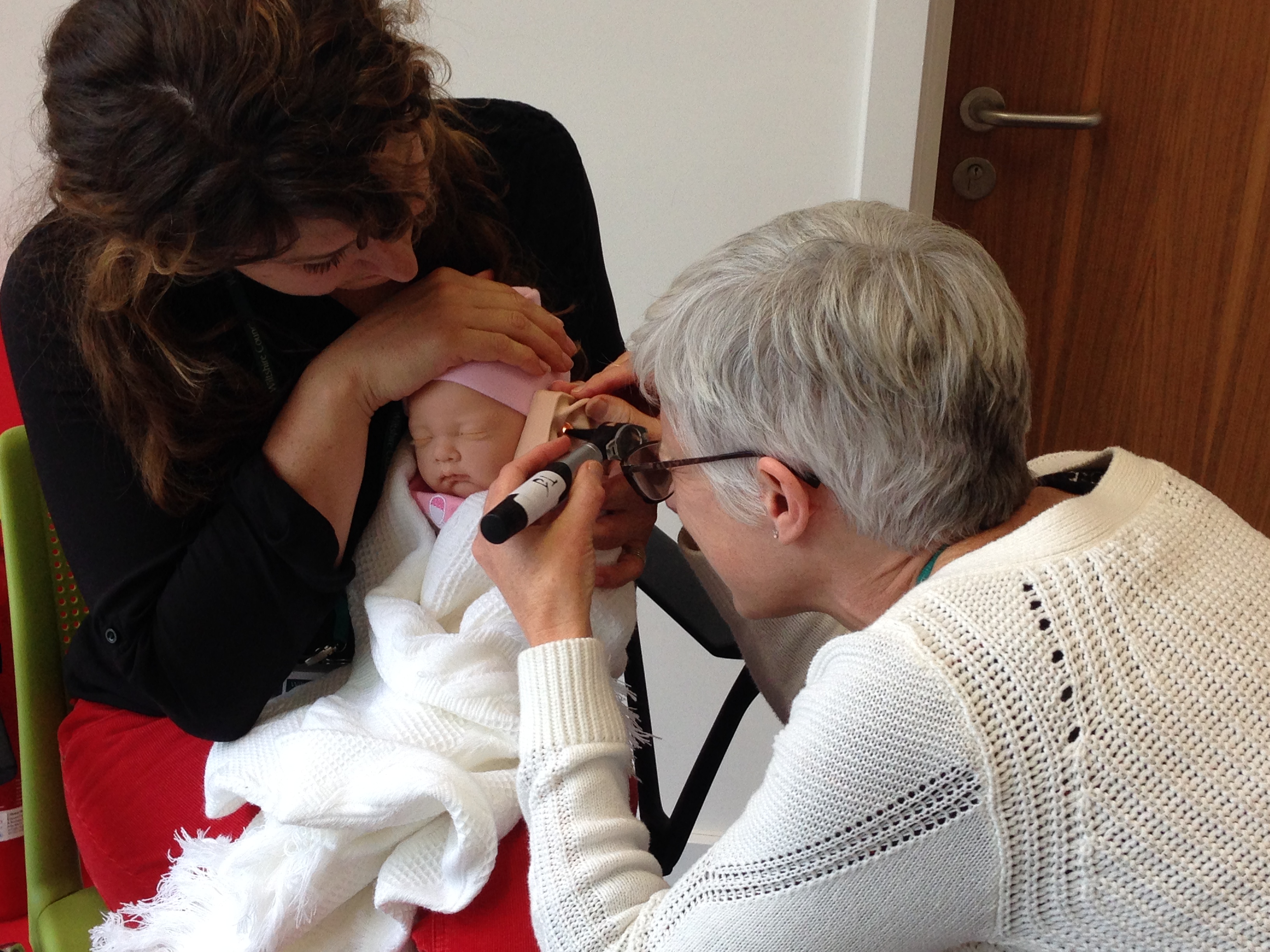
Otoscopy on a newborn baby

Masking is an ongoing lesson in Audiology.

As audiologists we all learn masking as part of our training. But the truth is, the real learning is in the experience of the various hearing losses we come across. Unfortunately for some, we’re not always in the position to reflect with another practitioner about some of the more complex hearing losses – so how can we really learn?

Masking is carried out as a result of the minimal interaural attenuation of 40dB when using headphones, 55dB when using inserts and 0dB when using a bone conductor. As a result, we follow certain rules to decide when masking should be carried out (BSA recommended procedure 2011) to ensure an accurate hearing test is obtained.

We probably know the rules by heart. But…

Why do we need to do Rule 3 and what is it’s relationship to Rule 1? What about central masking and the effect of conductive losses on masking? When are inserts more appropriate than headphones? When do we choose to mask bone conduction on both ears? What is effective masking? Even the most experienced of audiologists have to reflect on such questions when considering best practice.

A training course in masking in audiometry will enable audiologists to consider and understand why we need to mask, rather than just learning the rules. TJ Audiology Services uses audiometry simulation software to practice different scenarios, alongside interpretation of working audiograms. Audiology Courses are available in 2016 at Audiology House, London. Bring yourself up to speed, or start from scratch and benefit from having the time to ask all those questions and more in a safe learning environment and discussion with other colleagues.


BSA Recommended Procedure (2011): Pure-tone air-conduction and bone-conduction threshold audiometry with and without masking.


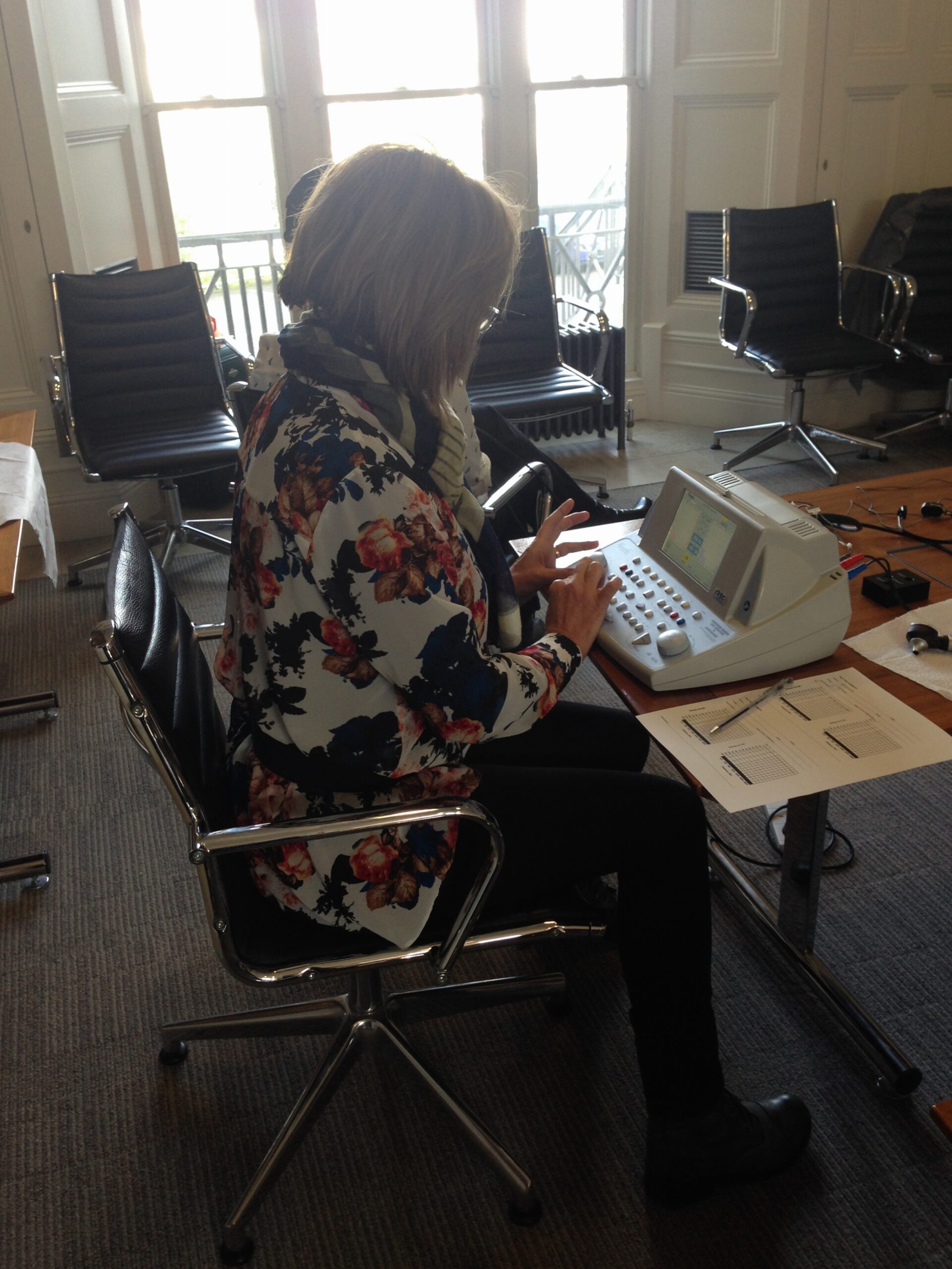
Audiometry and tympanometry for professionals who are not qualified audiologists

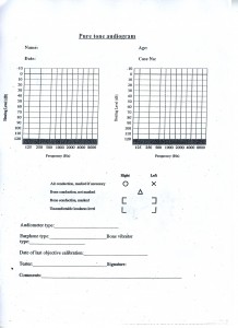
pure tone audiogram
pure tone audiogram

A hearing test (audiometry) measures the quietest sound and individual can hear, at least 50% of the time – known as hearing threshold level (HTL). An individual’s HTL at different frequencies is recorded in an audiogram for each ear, and this information can be used to categorise hearing as within normal range, or a hearing loss that can range from mild to profound. Otoscopy and tympanometry is used alongside audiometry to identify any abnormalities of the middle ear that may be affecting the hearing, e.g. perforation or glue ear.

In hospitals and hearing aid dispensing practices, audiometry and tympanometry is typically carried out by audiologists who are trained to undertake these procedures during their university course. In the UK, audiometry is carried out according the British Society of Audiology Recommended Procedure.

However, basic hearing tests and tympanometry can also be performed by GPs, occupational health nurses, audiometricians, assistant technical officers, teachers of the deaf and hearing aid assistants. The British Society of Audiology has a Practice Guidance Document for “Hearing assessment in general practice, schools and health clinics: guidelines for professionals who are not qualified audiologists”.

A British Society of Audiology accredited course in basic audiometry and tympanometry trains non-audiologists to undertake hearing tests in the field and interpret the results, according to minimum training criteria.

Are you going around the 2nd bend? How to make a good impression.

Good fitting custom earmoulds and hearing protection, ITE and CIC hearing aids can only be created from accurate impressions. The key is having the confidence to know when you’ve inserted your otostop deep enough down the canal, and to point the otostop in the direction of the eardrum. A good, deep impression means getting to the 2nd bend of the ear canal. Some tell-tale signs that you’ve reached the 2nd bend include a characteristic slip of the otostop into position which often coincides with slight resistance as it enters the bony portion of the canal. When you look on otoscopy, you will see that the otostop is framed by the cartilaginous portion of the canal.


All ear canals are very different from one another and taking good impressions takes practice. Patient/client safety is paramount and the British Society of Audiology has a recommended procedure for impression-taking in adults/children over 5 years of age and for children under 5 years of age.

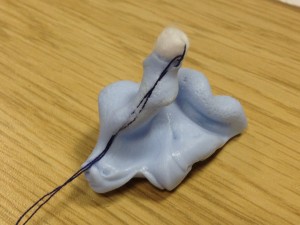
Second bend impression