Our courses offer an opportunity to gain experience and develop new skills in audiology. If you’re considering joining a TJ Audiology Training course we want to ensure that get the most out of it. Here are some tips to help you get the full benefit from your training with us:
Understand what you want to get from the course – We ask that you read the relevant pre-course information documents to understand what the course will cover and the key objectives. Approximately 2 weeks before your course we will send an email with further information documents and website links – we highly recommend that you take the time to read this information as it will help you to prepare for the course.
Ask Questions – We encourage all delegates to ask questions during the course. Tracy and Louise welcome questions and they will be able to answer your points, open them up as a discussion to other delegates or find out the answers after the course and get back to you.
Get Involved – Our courses are designed to be interactive and we want delegates to be involved. Our course trainers will ask the group questions or set tasks/practical assessments and we encourage you to get involved.
Implement your new skills and knowledge – The best way to develop your new skills is to practice them in your workplace whenever possible.
We will provide you with comprehensive training materials to help enhance the skills and knowledge that you acquire during the course. In addition to the information that we will send you, we encourage you to follow our Facebook page for updates and links, and keep up to date with our new posts here.
Otis – Audiology Training with Virtual Patients
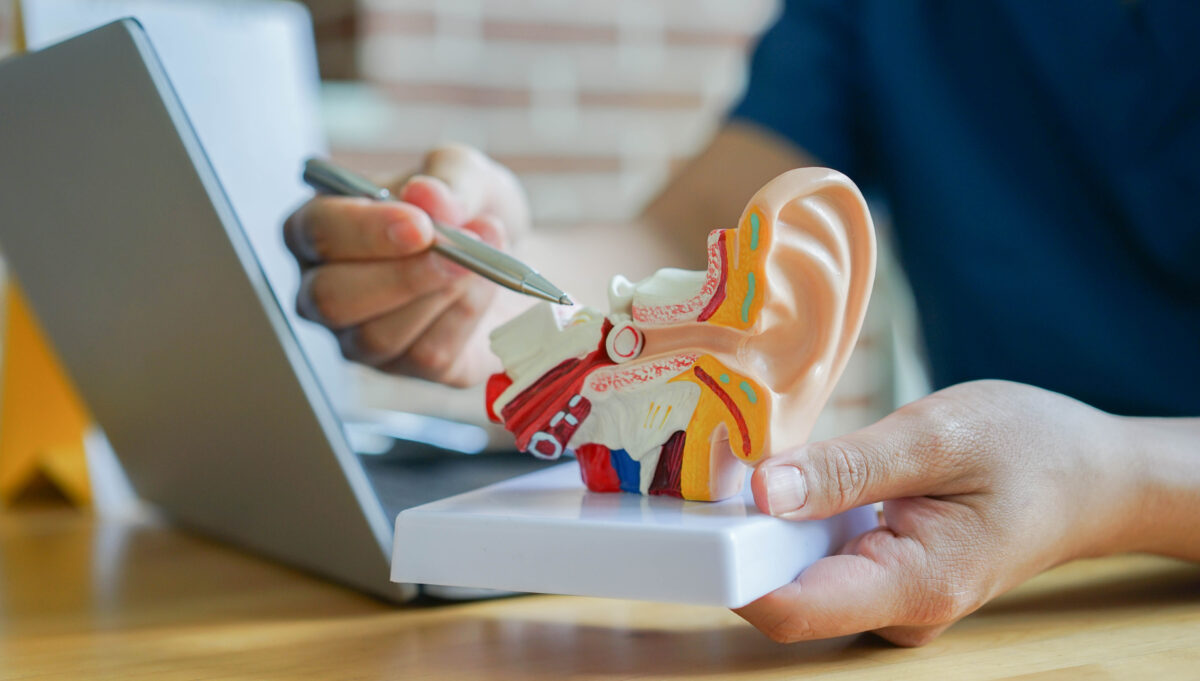
The structure and function of the ear and its role in hearing and balance
Cochlear Anatomy Video : Sam Webster