We are onto Part 3 already. This next block will be looking at possible audiograms for pathologies such as Otosclerosis, Meniere’s disease, ototoxic medication etc. Remember audiograms will differ depending where they are along the pathology pathway. Writing this I realised there is too much information to give on each topic; so, we will keep to one pathology for each of the upcoming blogs. As often these losses are progressive, I will try examples of early on the disease pathway and later on when the loss has progressed. Otosclerosis – FRCS ENT Exam Essentials is a video format that also helps explain the below.
Otosclerosis
Bone tissue in the body renews itself by replacing old tissue with new. In Otosclerosis regularly re-laid spongy bone, hardens abnormally and leads to fixation and this impairs the movement of the bone; most commonly the stapes. This affects the conduction of sound from the middle ear to the cochlea, causing a conductive hearing loss. Over 50% of people with otosclerosis, have a family history of the condition. Is an autosomal dominant mode of inheritance. Prevalence in women is twice that of men. We know that pregnancy can worsen the condition (we have yet to understand clearly why). We mostly see it in people of European origin, in people of Asian origin it is less common and even less so in people of African descent. Otosclerosis – StatPearls – NCBI Bookshelf. (Information for your client-Otosclerosis and Stapedotomy | ENT UK)
They will have the following presentation.
- Gradual decline in hearing usually seen to start in people in their 20-30’s however it is the most noticeable in their 30’s. It can start as early as 11 yrs of age to 45 yrs old. (earlier presentation in individuals with a family history)
- Conductive hearing loss with Carhart’s notch at 2KHz.
- Usually starts in one ear and then moves to the other. This loss may appear very gradually. Many people with otosclerosis first notice that they are unable to hear low-pitched sounds or can’t hear a whisper. Some people may also experience dizziness, balance problems, or tinnitus.
- Excellent speech discrimination scores (even with cochlear otosclerosis)
- Tympanometry wise, normal middle ear pressure and ear canal volume, static compliance is either normal or shows stiffness (0.3 or under)
- Absence of middle ear reflexes.
- Worsens with pregnancy/ is often first noticed during pregnancy
- Menopause, trauma, surgery have also been noted as aggravators of otosclerosis
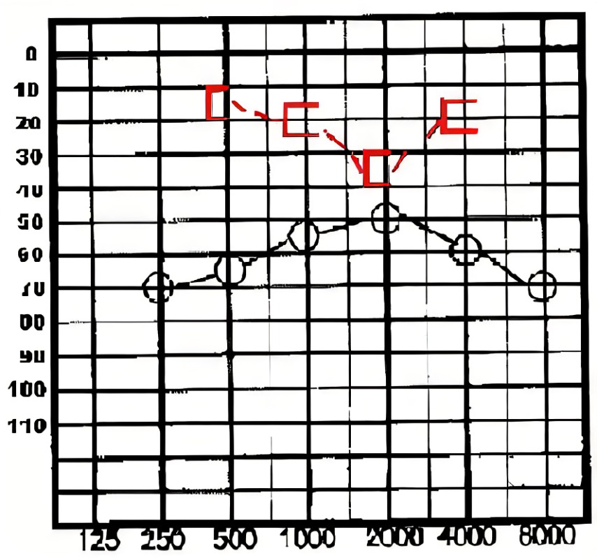
Audiogram, Otosclerosis. An audiogram showing bilateral mixed hearing loss in a patient with otosclerosis. The typical dip in bone conduction is also present.
Above you will see a dip at 2000hz we call this a Cahart’s notch. This is a mechanical artifact of testing, rather than being a true sensorineural hearing loss. The normal ossicular resonance in humans is around 2000 Hz, which is impaired by stapes fixation. If the client has surgery this notch may disappear.
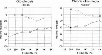
This is quite different to an audiogram for otitis media
Dr Chris de Souza ENT Specialist Audiological Investigation of Otosclerosis gives the below great explanation of the disease pathway for a client with otosclerosis
- Early stages
In the early stages of otosclerosis, audiograms show a conductive hearing loss.
- Late stages
As the disease progresses, the hearing loss worsens in the high frequencies and the air-bone gap widens. The audiogram changes from an upward sloping pattern to a flat pattern.
- Cochlear involvement
If the disease extends into the cochlea, a sensorineural hearing loss can develop. This is called cochlear otosclerosis.

This is an example on the right of advanced otosclerosis and is post stapes surgery
Intervention Options
Watch and Wait
People progress differently, so in some cases it is a watch and wait. Interesting to note that in women it may decline quicker with each subsequent pregnancy.
Hearing Aids
If there is a loss that can be aided then hearing aids will work well as it is a conduction of sound issue. This may also be the preferred treatment over surgery if the individual has another pathology on the other ear, such as an Acoustic Neuroma or Meniere’s. I have in the past indicated to my clients when progression of the otosclerosis is occurring (sensorineural component is progressing) and they may be getting to the stage where medical intervention will be less effective. This allows them to choose between staying with a hearing aid or going for surgery.
Surgery
The current standard medical treatment is a stapedectomy. A prosthesis is put in to replace the damaged bone. By removing the diseased bone and replacing it with a prosthesis this will allow the vibration of sound in the middle to work effectively again.
Patients with far advanced otosclerosis as discussed in the hearing aid section, and severe hearing loss, can still benefit from stapedectomy surgery but they will still need to use a hearing aid (as shown in the audiogram above). Otosclerosis – Vestibular Disorders Association